Periodontal disease: the most common complaint in the canine and feline oral cavities
Periodontal disease is the most common dental problem seen in dogs and cats. By contrast, the most common dental problem seen in people is tooth decay (cavitation), which rarely occurs in dogs (representing less than 10% of canine dental problems) and never occurs in cats. This means that when owners approach us wondering if their pet has tooth decay, the problem usually turns out to be periodontal disease. This form of disease attacks the gums and bone that support the teeth, and may ultimately result in tooth loss (Fig. 1). In small dogs, aggravation of periodontal disease can lead to swelling and perforation in the cheeks (Fig.2), or fractures of the jaw (Fig.3 and 4).
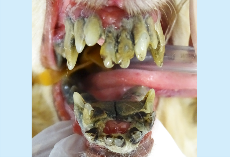
Figure 1: Severe periodontal disease
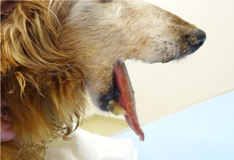
Figure 3: Mandibular fracture caused by periodontal disease
Figure 2: External dental fistula due to periapical lesion
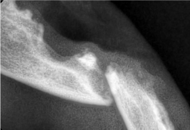
Figure 4: X-ray of mandibular fracture
Preventative measures should be taken to stop pets developing serious periodontal disease. And the necessary steps include home dental care and thorough examination of the oral cavity at an animal hospital. Findings of gingivitis or periodontitis may necessitate tartar removal or periodontal surgery.
Dental disease does not resolve naturally when left untreated. Please consult your veterinarian if you have any concerns about your pet’s teeth or oral health.
Although general anesthesia is sometimes cautioned against for geriatric pets, old age should not be seen as medical condition in itself. The highly trained veterinary anesthesiologists at our hospital know how to anesthetize your pet in the safest possible way. Before undergoing surgery, each pet is examined thoroughly to make sure it can be anesthetized safely. Please do not hesitate to tell us if you have any worries or concerns.
Oral tumors are also common in older dogs and cats
The following symptoms may be signs of oral tumors: halitosis; difficulty eating hard food; weight loss or loss of appetite; and bleeding gums. Oral tumors are often malignant and can be life-threatening. Therefore, it is crucial to find, diagnose and properly treat them as early as possible. For older dogs and cats, the oral cavity should be examined regularly, and any concerns should be discussed with your veterinarian as early as possible. For veterinary practitioners, please do not hesitate to consult us if you have any questions or doubts about diagnosis or treatment.
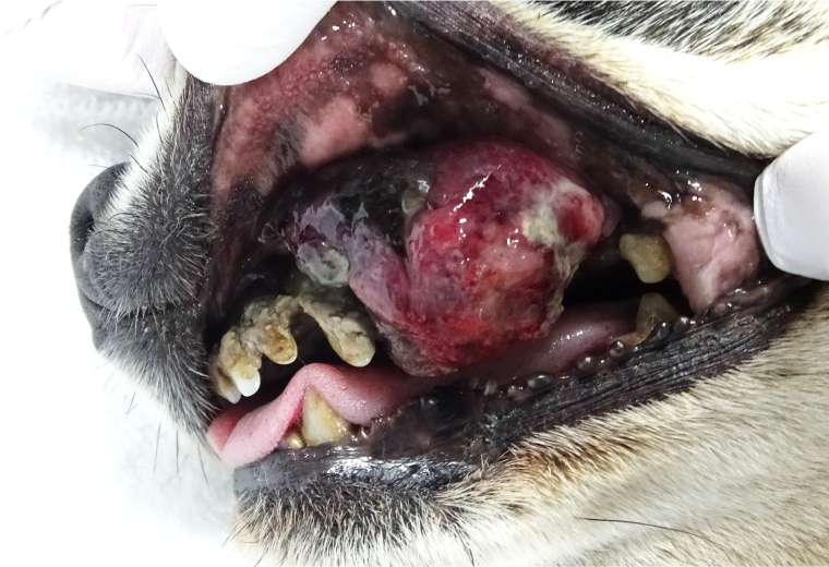
Figure 5: Malignant melanoma (example of an oral cavity tumor)
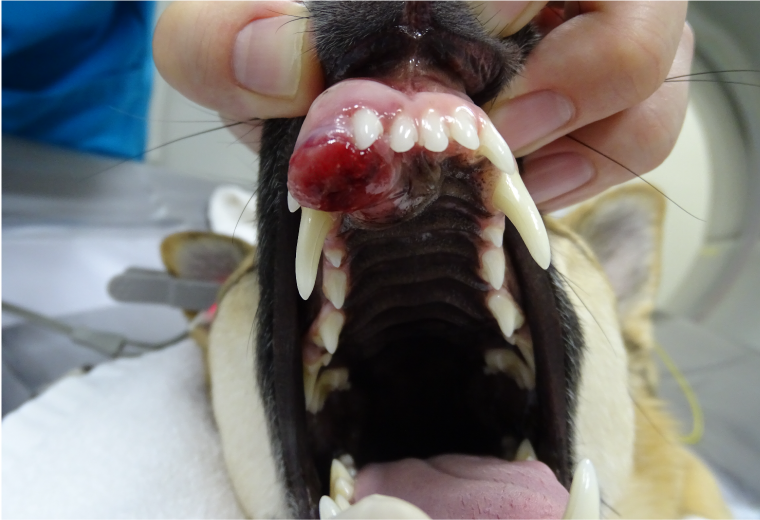
Figure 6: Tumor in the oral cavity
In both cases, the patients underwent tumor resection, which included a partial maxillectomy, with satisfactory outcomes.
About oral surgery
When undergoing oral surgery, animals are put under general anesthesia to prevent them from suffering pain and anxiety.
Each animal undergoing anesthesia is examined to determine its overall condition and the least stressful anesthetic that can be used. The animal’s blood pressure, heart rate, body temperature, and other vital signs are carefully monitored throughout the operation.
We perform such procedures as dental scaling (teeth and bone) and aspiration of the oral cavity using a veterinary dental unit equipped with a scaler, digital dental X-rays, and CT scans as necessary.
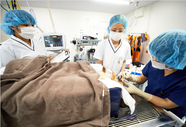
Figure 7: Surgery
Anesthesia monitoring during surgery
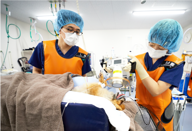
Figure 8: Digital dental X-ray
Dental radiography is an essential tool for diagnosing and making treatment plans for oral conditions.
Below, we outline the wide range of treatments that can be offered according the patient’s symptoms and condition. In all cases we proceed with the best course of treatment for the patients fully discussing the case with their owners.
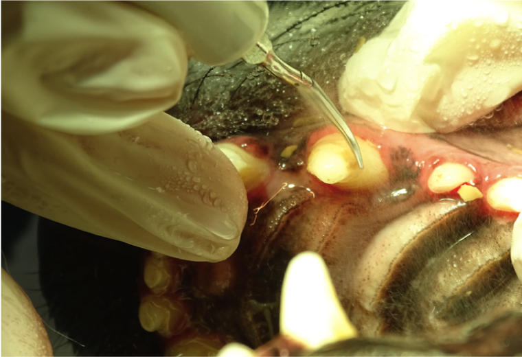
Root canal therapy for fractured teeth (broken teeth)
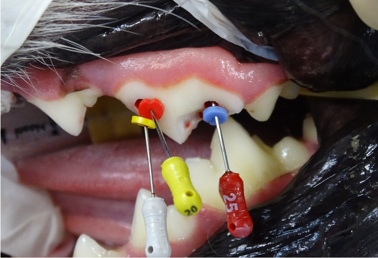
Figure 9: Root canal therapy (root canal filling)
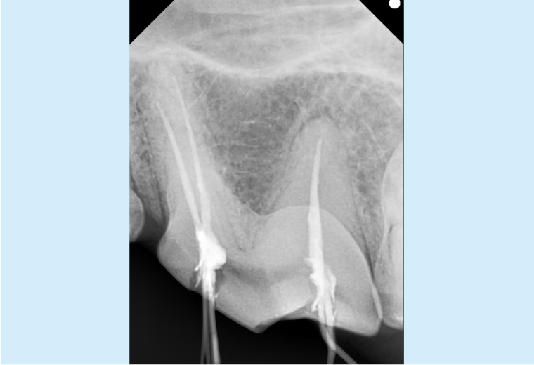
Figure 10: Digital dental X-ray of filling
We remove infected pulp from the root canal, clean and sterilize the canal, and replace the removed tissue with dental material.
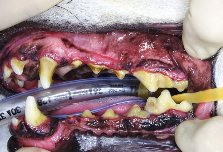
Orthodontics
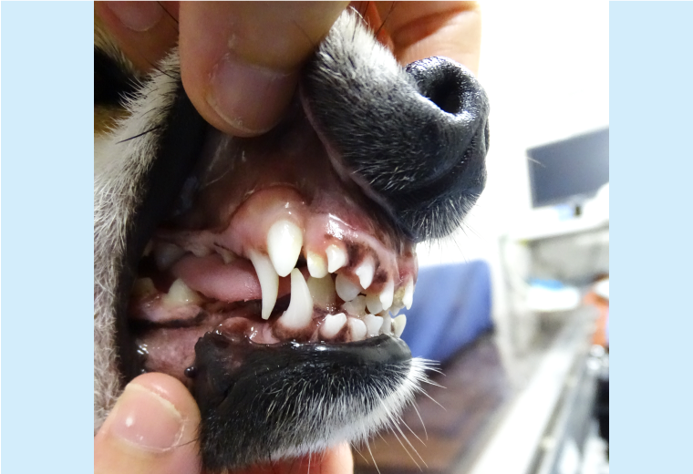
Figure 11: Before surgery
This dog has not shed many its primary teeth, and its canine tooth on the lower jaw is angled inward.
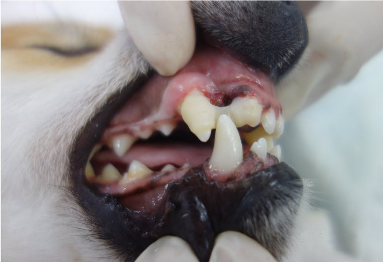
Figure 12: After surgery
Wee extracted the retained primary teeth, and corrected the canine tooth on the lower jaw, to achieve a functional bite.
Mandibulectomy and Maxillectomy
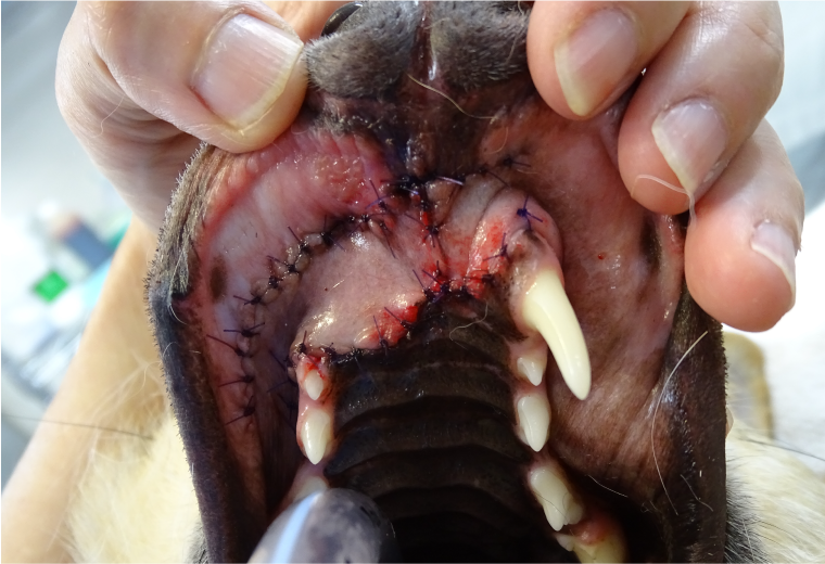
Figure 13: After maxillary resection
The tumor was completely removed, leaving the dog with its appearance little altered and its ability to eat unaffected.
Mandibular reconstruction resulted from fracture
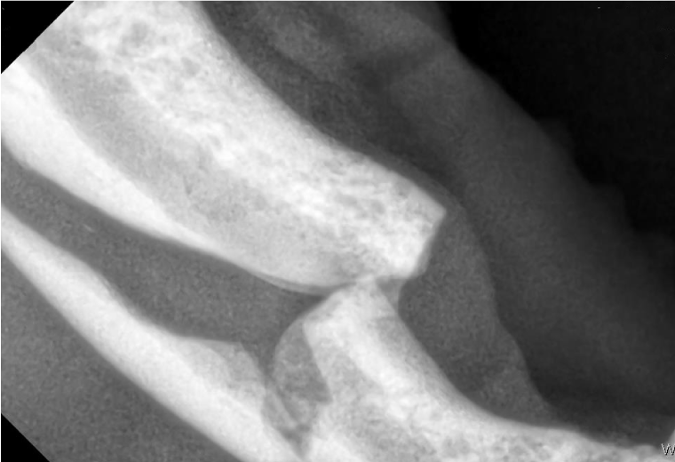
Figure 14: X-ray of mandibular fracture
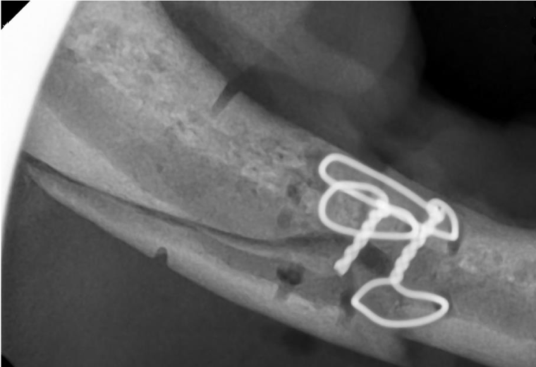
Figure 15: Wire-assisted fracture reduction
Vital pulpotomy of canine tooth
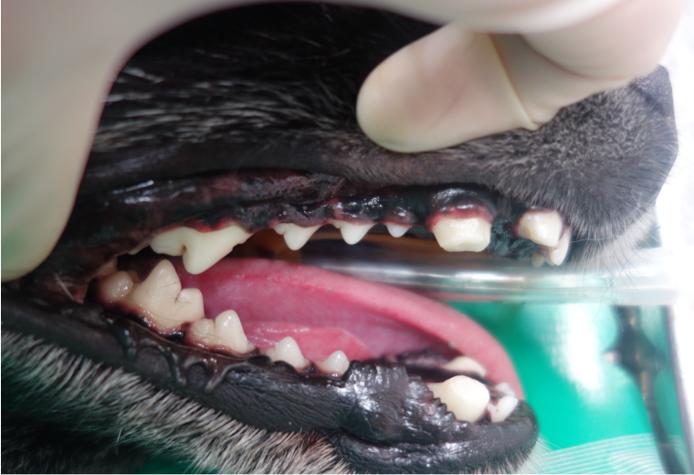 Figure 16: Vital pulpotomy (canine tooth resection)
This dog underwent dental disarming (crown amputation of the upper and the lower canine teeth) as it had frequently bitten people. This procedure involved nerve management for the re-sectioned tooth surface and capping with composite resin.
Complete premolar and molar extract to treat a cat with gingivostomatitis
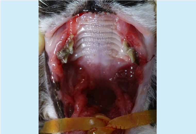
Figure 17: Before surgical treatment for gingivostomatitis
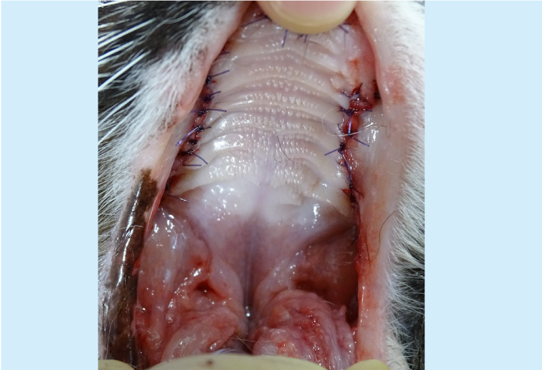
Figure 18: After surgical healing
The rubefaction and swelling in the oral cavity subsided and the pain was alleviated.
Procedure for plaque and tartar removal at KUVTH
1. Examination and evaluation: The patient is placed under general anesthesia, and the veterinarian then checks the teeth, periodontal pockets, gums, and supporting bone.
2. Scaling: Visible tartar is removed from the tooth surface using an ultrasonic dental scaler while rinsing with cold water.
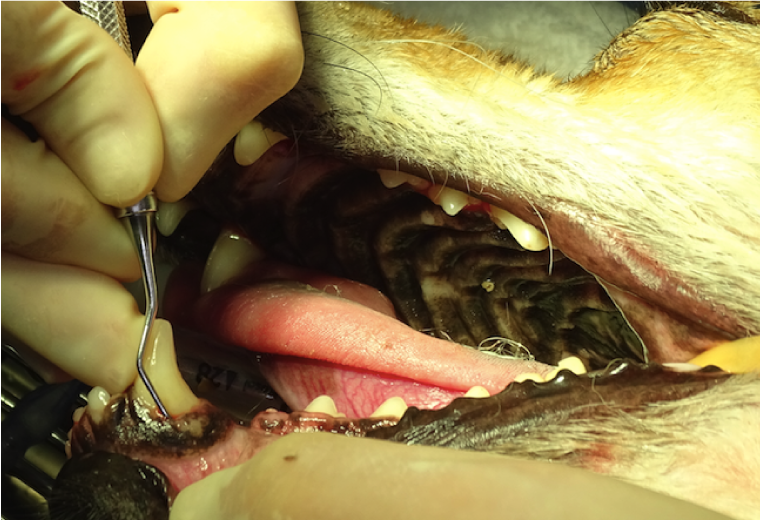
3. Curettage/Root planing: Tartar is removed from the periodontal pockets using curettes, and the tooth root surface is smoothed.

4. Polishing: The tooth surface is polished with a coarse, green abrasive agent and a polishing brush.
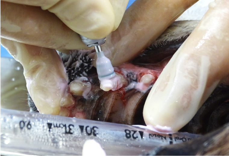
5. Finishing: The tooth surface is subject to a finishing polish with a fine, pink abrasive agent and a rubber cup to prevent any recurrence of plaque and tartar accumulation.
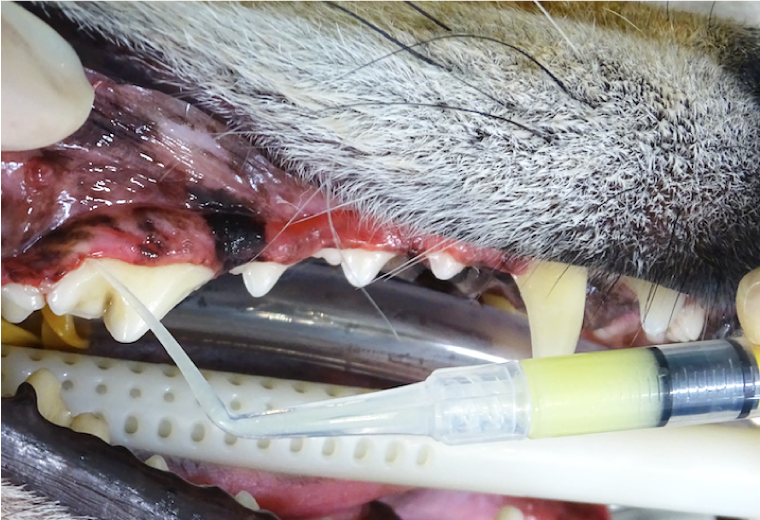
6. After scaling, a doxycycline/slow-release dental antibiotic ointment is applied to any inflamed areas and deep pockets.
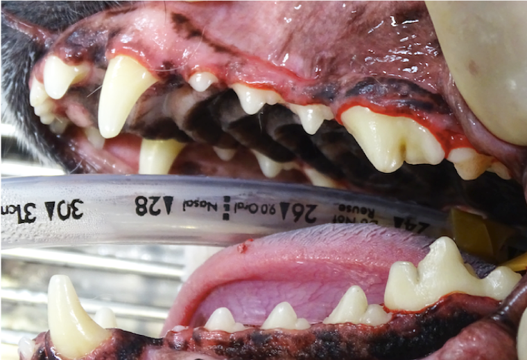
7. Scaling is now completed. If there are deep periodontal pockets, dental X-rays will be taken to check for dissolution of the bone surrounding the tooth and the presence or absence of periapical lesions (inflammation and bone resorption around the tooth root tip), which may then necessitate tooth extraction or some other further treatment.
Dental care often requires specialized equipment, with X-rays equipment being a prime example. If you notice your pet displaying any symptoms or abnormal signs, please contact us as soon as possible.