Diagnostic Imaging (Radiology)
The KUVTH Diagnostic Imaging Unit has two mottos: “We do our utmost to treat animals compassionately” and “we diagnose accurately”.
Recent years have seen great advances in diagnostic techniques at animal hospitals. For example, X-radiography and ultrasonography have now become standard procedures in many veterinary hospitals. Animals cannot explain their symptoms, or where pain is located, or whether a drug is providing relief. When veterinarians determine something is wrong with an animal, they have to do so based on that animal’s symptoms while studying its expression.
This explains why myriad medical imaging technology and equipment are even more important for veterinary than human medicine. The Diagnostic Imaging Unit at KUVTH utilizes a full complement of modern imaging modalities, including digital and computed radiography (X-ray), endoscopy, ultrasonography, X-ray, computed tomography (CT), and magnetic resonance imaging (MRI). We also possess portable X-ray, endoscopy, and ultrasound systems which allows us to offer field veterinary examinations of large animals. We have a 16-slice helical CT scanner, and more than 300 cases are imaged every year. At KUVTH, we constantly strive for animal-friendly imaging, and anesthesia-free CT scans have been part of our hospital’s approach since an early point in our history. Now, more than 80% of the procedures for CT scans are performed without anesthesia.
In addition, our 3-tesla MRI system is state-of-the-art technology enabling more accurate diagnoses. The 3-tesla MRI system enables detection of lesions such as cerebral infarction which were difficult to detect with conventional MRI.
As a major distinction of KUVTH, this equipment can be utilized for large animals—cattle and horses. Our CT system has been specially modified to hold large animals weighing up to nearly one ton. We also contribute to the local industry through our veterinary services offering examinations and testing services.
With optimal use of this equipment, we strive to deliver timely, accurate and skillful diagnostic imaging service for our patients.
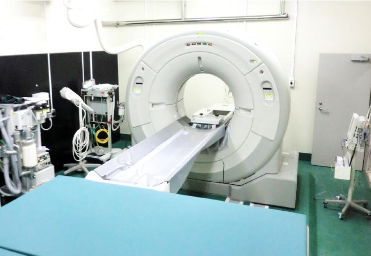
CT
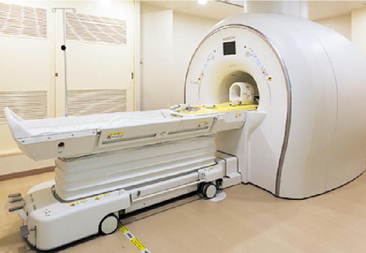
MRI
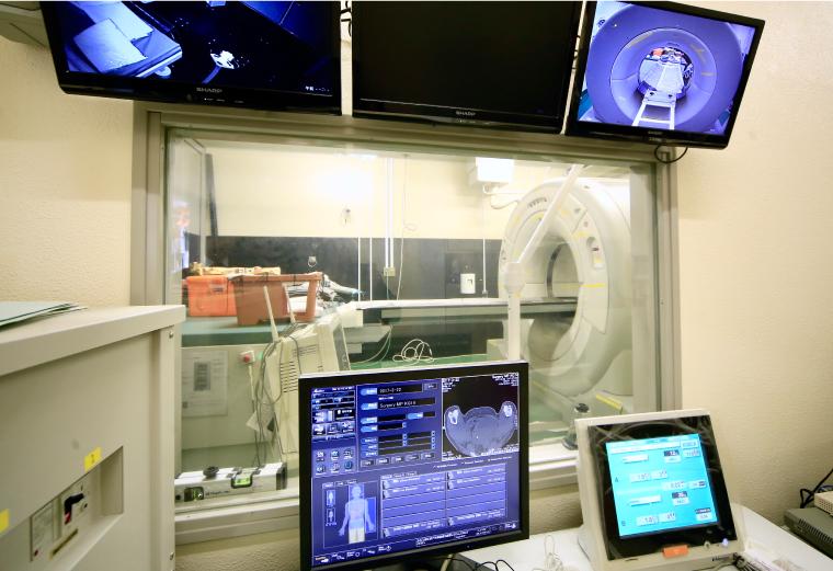
Control Room
Clinical Pathology
Animal hospitals have to make comprehensive diagnoses based on a range of medical examinations because animal patients do not give their doctors any verbal information. Clinical pathology—the tests performed in the laboratory—is thus a crucial source of information.
Regular blood tests encompass complete blood cell counts (CBCs) and blood chemistry. CBCs quantify the different cell types in the blood such as white blood cells, red blood cells, and platelets, and can be useful for diagnosing anemia and other conditions and infections. Blood chemistry tests quantify blood components, and can be useful for diagnosing diseases and identifying abnormalities in the liver, kidneys and other organs. Urinalysis and fecal tests can also play an important role in diagnosing disease. For example, urinalysis can identify renal diseases in cases where there have been no abnormalities in blood tests. Other crucial tests are cerebrospinal fluid (CSF) analysis, which assists in the diagnosis of neurological conditions, and synovial (joint) fluid analysis, which assists in the diagnosis of arthritis. Another area of clinical pathology receiving much attention recently is cytodiagnosis.
Cytodiagnosis can be simply described as microscopic examination of cells. Cells can be obtained in a variety of ways ranging from fine needle aspiration to centrifugation of thoracic and abdominal fluids, and they are examined microscopically as part of diagnosing cancer and infectious diseases. The examination of cells subject to special staining enables us to diagnose diseases that may be undetectable in regular tests.
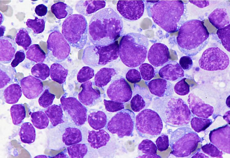
Cytodiagnostic investigation of a cat with generalized enlargement of the lymph nodes; lymphoma was suspected.
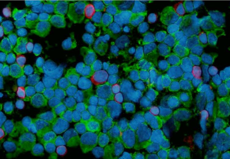
Special staining was applied for the same cat (as in the image on the left); B-cell lymphoma was diagnosed (and the diagnosis was substantiated with genetic testing). Blue stain: nucleus, Green stain: B-cells (CD79α), Red stain: T-cells (CD3)